バイオ3Dプリンタでヒトの心臓作製実現に近づく研究成果【カーネギーメロン大学】

3Dプリンタを使って、心臓の各部分をコラーゲンから作る研究成果がカーネギーメロン大学より発表されました。
3D bioprinting of collagen to rebuild components of the human heart
論文はサイエンス誌(2019年8月号)に掲載されました。
まず、最大のポイントは、
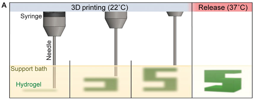
コラーゲンをニードルから押し出しても、受け止める何かがなければ、下のように水たまりになってしまいます。これでは立体臓器をつくれませんよね。

ほかのポイントは、
コラーゲンを積層するためにFRESHという技術を使っていること
今までにない20μmという解像度を実現していること
血管だけでなく、心臓、心室、三尖弁を再現していること
この研究のキーパーソンの1人は、Adam W. Feinberg氏。
出典:カーネギーメロン大学
同氏は、今回の研究成果が出る4年前の2015年に、同じ技術を使って血管を3Dプリントした論文を発表しています。
👇これは当時の論文内容を紹介する動画です。
2015年の論文(Three-dimensional printing of complex biological structures by freeform reversible embedding of suspended hydrogels)に、FRESH法についてもう少し詳しく書かれていました。
FRESHとは、freeform reversible embedding of suspended hydrogelsの略です。
下記はAdam W. Feinberg氏による2015年の論文からの引用です。
The key innovation in FRESH is deposition and embedding of the hydrogel(s) being printed within a second hydrogel support bath that maintains the intended structure during the print process and significantly improves print fidelity
The support bath is composed of gelatin microparticles that act like a Bingham plastic during the print process, behaving as a rigid body at low shear stresses but flowing as a viscous fluid at higher shear stresses. This means that, as a needle-like nozzle moves through the bath, there is little mechanical resistance, yet the hydrogel being extruded out of the nozzle and deposited within the bath is held in place.
Once the entire 3D structure is FRESH printed, the temperature is raised to a cell-friendly 37°C, causing the gelatin support bath to melt in a nondestructive manner.
上記内容について、
FRESHを用いるとなぜ、流動体のコラーゲンの変形を防げるか?にからめてまとめると、次の4点になります。
出典:Three-dimensional printing of complex biological structures by freeform reversible embedding of suspended hydrogels
①支持槽(Support bath)はニードルから押し出されたコラーゲンを支えることができる
②そのため、流動化しやすいコラーゲンでも、適所で凝固させることができる
③支持槽はバターのような性質をもつ。つまり、力を加えないときは硬く、力を加えると流動化する
④③の性質により、ニードルが支持槽の中を移動するときに受ける抵抗は小さく、押し出されたコラーゲンは適所に配置される
お菓子作りをする人にはおなじみですが、バターに力を加えて混ぜていくと、ある時からとても滑らかになりますよね。

このように、力を加える前は硬いのに、一定の力を加えると柔らかくなる性質の物体を、ビンガム流体といいます。
論文で使っているハイドロゲル(支持槽を構成するもの)は、ビンガム流体のようにふるまうわけですね。
出典:Three-dimensional printing of complex biological structures by freeform reversible embedding of suspended hydrogels
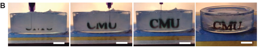
上記写真は、「CMU」という文字を3Dプリントしているところ。
支持槽を構成するハイドロゲルは、37℃になると溶けます。
プリント完了後に温度を上げると、写真の一番右側の状態のように、支えていたゲルが液体になるため、「CMU」が浮かんできます。
※ちなみにCMUは、カーネギーメロン大学を意味しています。
映像はこちらのページにある「Movie S1」という動画で確認できます。
上記でかいたことが、FRESH(freeform reversible embedding of suspended hydrogelsの略)という3Dプリンティング技術を指すようです。
FRESHによる3Dプリンティングのことを、英語では「FRESH-print」というようです。
このFRESHを使った3Dバイオプリンティングに関する最初の論文は、おそらく冒頭で触れた下記論文です。
今回の論文も、2015年の上記論文も、キーパーソンの1人はAdam W. Feinberg氏です。
今回注目されるのは、主に次の点です。
コラーゲンを使って毛細血管から三尖弁、心室、心臓全体を3Dプリントできること
流動体のため積層造形の難しかったコラーゲンを使えること
血管だけでなくヒトの心臓をプリントする可能性を示したこと
患者のMRIデータから患者特有の心臓を再現できること
今回の論文では、FRESHを改良して、急速にpHを変化させるアプロ―チを採用しています。
これによって、化学修飾したコラーゲンを使うことなく、機械的性質を強化することができ、従来のたるみやすい問題をクリアできているようです。
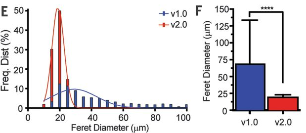
FRESHバージョン1とFRESHバージョン2の違い
今回の論文で使われているFRESHは改良版のバージョン2。
バージョン1との大きな違いは、解像度です。
バージョン1ではコラーゲンフィラメントの直径が250μmであったのに対し、バージョン2では、直径20~200μmの解像度を実現しています。
In contrast, FRESH v2.0 improves the resolution by an order of magnitude, with collagen filaments reliably printed from 200 μm down to 20 μm in diameter
3D bioprinting of collagen to rebuild components of the human heartより引用
※今回使われている方式は、押し出していくディスペンシング方式ですが、ディスペンシングでは限界と言われていた高解像度をどのように実現しているのか、装置に関する説明は確認できませんでした。私の理解不足かもしれません。
ちなみに、粒子は不規則な形状をしているため、日常で使う「直径」の定義をそのまま適用できませんよね。
論文ではフェレー径(Feret Diameter)を使用しています。
👇フェレー径とは、平行線で挟んだときの距離(幅)のことです。
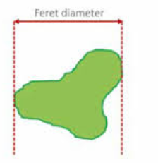
出典:Part icle size analysis of non-spherical part icles
確かにバージョン1より改良版のほうが、フェレー径が小さくなっていますね。
出典:3D bioprinting of collagen to rebuild components of the human heart
2019年に報告されたほかの心臓バイオプリンティング
今年、2019年に注目された別の論文に、テルアビブ大学から発表された心臓の3Dプリンティングがありましたね。このブログでも紹介しました。
成功したのはまだ小さなサイズでしたが、血管と心臓を同時に3Dプリントするのが特徴でした。
テルアビブ大がハイドロゲルとして使ったのは、患者の大網由来の細胞を脱細胞化したECM。
今回のカーネギーメロン大学の論文にも、テルアビブ大の研究成果が触れられていますが、「構造的・機能的な解析がなされていない」と指摘されています。
Recently, Dvir and colleagues 3D-printed a decellularized ECM hydrogel into a heart-like model and showed that human cardiomyocytes and endothelial cells could be integrated into the print and were present as spherical nonaligned cells after 1 day in culture (8). However, no further structural or functional analysis was performed.
3D bioprinting of collagen to rebuild components of the human heartより引用
【参考】テルアビブ大の論文は下記よりどうぞ。
3D Printing of Personalized Thick and Perfusable Cardiac Patches and Hearts
今後、バイオプリンティングの研究においては、心臓を中心とした研究が活発になっていく気がします。
今回、カーネギーメロン大学でプリントした心臓は、新生児の心臓と同じサイズです(下記写真)。ヒトにはまだ移植されていません。
Finally, to demonstrate organ-scale FRESH v2.0 printing capabilities and the potential to engineer larger scaffolds, we printed a neonatal-scale human heart from collagen.
3D bioprinting of collagen to rebuild components of the human heartより引用
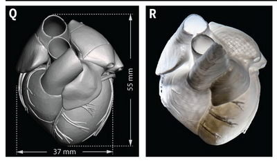
出典:3D bioprinting of collagen to rebuild components of the human heart
移植臓器が足りないという喫緊の課題が解決される日は近いでしょうか。
成長とともに心臓が大きくなるスピードの速い小児患者にとっても、目が離せない研究です。
英語ニュースでは、Being able to 3D print a full-sized adult human heart just got much closerとタイトルがつけられていました。
バイオプリンティングについては、まだ勉強不足です。今後はもっと深掘りできる記事を目指します。
※アイキャッチ画像の出典:カーネギーメロン大学
【参考】
今回の論文:3D bioprinting of collagen to rebuild components of the human heart
Part icle size analysis of non-spherical part icles